Dominic E. Nathan, MS;1�2 Michelle J. Johnson, PhD;1�3* John R. McGuire, MD³
¹Department of Biomedical Engineering, Marquette University, Milwaukee, WI;
²Rehabilitation Robotic Research Design Laboratory, Clement J. Zablocki Department of Veterans Affairs Medical Center, Milwaukee, WI;
³Department of Physical Medicine and Rehabilitation, Medical College of Wisconsin, Milwaukee, WI
Abstract
Hand and arm impairment is common after stroke. Robotic stroke therapy will be more effective if hand and upper-arm training is integrated to help users practice reaching and grasping tasks. This article presents the design, development, and validation of a low-cost, functional electrical stimulation grasp-assistive glove for use with task-oriented robotic stroke therapy. Our glove measures grasp aperture while a user completes simple-to-complex real-life activities, and when combined with an integrated functional electrical stimulator, it assists in hand opening and closing. A key function is a new grasp-aperture prediction model, which uses the position of the end-effectors of two planar robots to define the distance between the thumb and index finger. We validated the accuracy and repeatability of the glove and its capability to assist in grasping. Results from five nondisabled subjects indicated that the glove is accurate and repeatable for both static hand-open and -closed tasks when compared with goniometric measures and for dynamic reach-to-grasp tasks when compared with motion analysis measures. Results from five subjects with stroke showed that with the glove, they could open their hands but without it could not. We present a glove that is a low-cost solution for in vivo grasp measurement and assistance.
Key words: functional electrical stimulation, grasp-assistive device, hand therapy, motion analysis, reach to grasp, rehabilitation, robotic stroke therapy, stroke, upper limb, validation.
Abbreviations: 3-D = three-dimensional, ADL = activity of daily living, ADLER = Activities of Daily Living Exercise Robot, ANOVA = analysis of variance, DOF = degree of freedom, FES = functional electrical stimulation, GUI = graphical user interface, MCP = metacarpophalangeal, MMT = manual muscle test, PIP = proximal interphalangeal, SD = standard deviation, UL Model = Bilateral Upper-Limb Kinematic Model.
Introduction
More than 5 million people in the United States are dealing with disabilities related to stroke, the leading cause of disability among adults in the country [1]. These disabilities affect patients� ability to accomplish real-life activities of daily living (ADLs), such as drinking and eating. These ADLs often involve critical submovements, including reaching and/or grasping. About 500,000 new strokes occur each year, leaving about 66 percent of these patients with residual upper-limb motor impairments and about 50 percent without functional independence [1]. New stroke rehabilitation insights suggest that repetition, intensity, motivation, and skilled task-oriented practice are key to poststroke functional recovery processes and may lead to use-dependent functional reorganization [2�5]. Our ultimate goal is for patients with stroke to regain the ability to accomplish a variety of skilled tasks involving reaching and grasping using both the arm and hand.
Typically, robotic therapy environments designed for stroke rehabilitation focus on retraining motor control using only reaching tasks, without focusing explicitly on tasks involving the hand [6�11]. Recently, a conscious effort has existed in the rehabilitation robotics field to include the hand in robotic therapy. As a result, one such development is the Activities of Daily Living Exercise Robot (ADLER), a robotic therapy environment focused on using reaching and grasping activities to retrain motor and ADL function (Figure 1) [11]. Preliminary experience with this functional robot indicates that without an assistive hand device coupled with the reach assistance provided by the robot, patients with stroke who are lowfunctioning or lack hand function would be limited to only assistive or resistive reaching activities. To remedy this, we set two main goals. The first was to integrate a low-cost grasp-assistive glove into ADLER that would enable hand therapy, and the second was to measure grasp aperture as a user completes simple-to-complex ADLs with and without the ADLER.
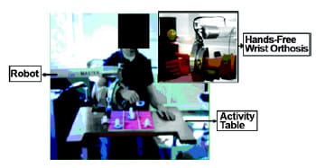
Figure 1. Activities of Daily Living Exercise Robot setup indicates robot
setup for desktop activities such as game playing. System uses MOOG FCS HapticMaster
(EST; Kaiserslavtern, Germany) with custom hand attachment to accommodate hands-free interactions.
From the literature, we identified current commercially available or research-based grasp-assistive devices and hand-measurement tools. We explored whether these commercially available or current developmental systems could be integreated with ADLER and benchmarked them to determine key requirements. Current commercially available functional electrical stimulation (FES) systems such as the NESS H200 (Bioness Inc; Valencia, California) are secured to the forearm and can generate hand opening and closing but have limited application for fine manipulation tasks, specifically the grasping of small or circular-type objects. Mechanical hand-opening devices such as the SaeboFlex (Saebo Inc; Charlotte, North Carolina) and the CyberGrasp (Immersion Corp; San Jose, California) facilitate impaired hand opening using mechanical actuators of springs and pulleys. These devices often require extensive custom-fitting to patients, and a large and somewhat bulky profile prevents easy assimilation with functional tasks while the patient uses the ADLER.
Orthotic robotic devices developed for limb therapy (such as Rutgers Hand Master II-ND Force-Feedback Glove [12], the HWARD robotic hand-therapy device [13], the finger therapy robot at the Rehabilitation Institute of Chicago [Chicago, Illinois] [14], the GENTLE/G hand robotic therapy system developed at the University of Reading [Reading, United Kingdom] [15], and cabledriven finger therapy robots [16]) have been successful in providing grasp assistance and hand opening. However, this success is at the cost of a high degree of complexity in use, attachment, and construction. Since our goals are ADL func
tion, hand opening, finger extension, and support for real-life activities, we found too many of these devices were limited in assisting with real-world object and task therapy.
In examining current commercial options for measuring finger movements, we decided that sensorized gloves show promise and can measure finger joint angles and grasp aperture. A majority of these glove systems is geared toward the virtual reality market. Systems such as the 5DT Data Glove Series (Fifth Dimension Technologies, Inc; Irvine, California), the CyberGlove (Immersion Corp; San Jose, California), and ShapeHand (Measurand Inc; Fredericton, Canada) use between 5 and 22 sensors for measurements. Capable of providing information such as finger joint angle information and hand posture, these systems are often very expensive (>$1,000 a unit) and hard for patients with stroke to don and doff. Lowercost gaming systems such as the P5 glove (Essential Reality; New York, New York) provide gross grasp information but are unsuitable for accurate hand-opening or -closing measurements. The nature of the sensors employed and the large variability in the response accuracy of the glove prevent accurate hand-posture measurements.
The literature provided us with insight into features such as sensor number and placement, materials used, interface options, software, power supply consideration, and functionality that could influence glove design and data collection and provide grasp assistance. This article presents a low-cost glove to measure grasp aperture and facilitate hand opening and closing during task-oriented robotic therapy. To determine the reliability and validity of the glove as a tool to measure hand opening and closing, we compared results obtained from our glove with those obtained from gold standard measurement tools used during static and dynamic tasks. Results from five nondisabled subjects showed that our glove is accurate and repeatable for both static open and closed tasks when compared with goniometric measures and for dynamic reach-to-grasp tasks when compared with motion analysis measures. We also assessed assistive capability that is realized using a modified functional electrical muscle stimulator integrated with the glove. Results from five subjects with stroke showed that with glove assistance, subjects were able to open their hands but they were unable to without the glove. Overall results indicate that the glove can measure in vivo grasp aperture during functional tasks and may be used in robotic therapy.
Methods
Glove System Requirements
We centered the design of our FES grasp glove on a set of core requirements and analyzed each phase of the design process to ensure that these core requirements were adequately met [17]. The core requirements are outlined as follows:
- Integration with the ADLER system (Figure 1). The glove must be able to interface with the current ADLER system and not interfere with therapy-task performance. It must be portable and be usable with and without the ADLER.
- Comfort and durability. The glove must be minimally invasive, be lightweight, and not interfere with tasks performed by the subject. It must also be comfortable during use and adaptable to various hand sizes. Finally, it must be easy to don and doff, especially for individuals with reduced hand and finger range of motion.
- Function. The glove must be able to accurately predict grasp aperture of the hand and determine the grasp preshaping and release stages during a functional task using a real-world object. Grasp aperture is derived from the joint angles of the fingers. The glove should enable a more “natural” interaction with the subject and the object that is being manipulated.
- Low cost. The glove must be low in cost to both the research team and subject.
To meet these requirements, we developed the FES grasp glove. This glove system consists of five main parts: sensorized glove; grasp prediction model; muscle stimulation unit; custom data acquisition, controller, and processing programs; and controller circuit.
Grasp Prediction Model
The glove design is based on a new robot model for predicting grasp aperture as dictated by the position of the index finger and thumb (Figure 2). A pair of two-link robots connected to a rigid base represents these two digits. This grasp model assumes several key points. First, that functional grasp, i.e., hand opening and preshaping, for various functional tasks can be accurately predicted based on the aperture relationship of the index finger and thumb. The assumption is from observations of digit utilization during the main types of functional grasping, namely the tripod, pinch, and power functional grasp [18�22]. In our understanding of the roles of the fingers during grasp, the thumb and index finger are always involved in performing a major part of the manipulation. Second, for the index finger, the distal phalange and interphalangeal segments move as a unit and are considered a single rigid body. Third, the metacarpophalangeal (MCP) joint for the thumb is constrained to a single degree of freedom (DOF) revolute-type joint that only accounts for flexion and extension.
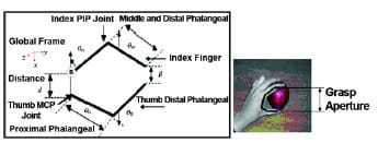
Figure 2. Proposed grasp model showing grasp aperture (β) as dictated by distance (d)
between tip of index finger and thumb. Hand shows application concept for model.
MCP = metacarpophalangeal, PIP = proximal interphalangeal, θ = joint angles, B = base frame.
The novel 4-DOF planar robot consists of four revolute joints, each one representing the proximal interphalangeal (PIP) and MCP joints of the thumb and index finger (Figure 2). We designated the MCP joint of the index finger as the base for the model marked B in the figure. The distance between the tips of the robot end effectors predicts the grasp aperture β, as dictated by the thumb and index finger, by using the joint-angle information of the PIP and MCP joints of these two digits. We obtained the Cartesian positions of the thumb (xthumb, ythumb) and index finger (xindex, yindex) from the joint angles and the lengths of the digits, as denoted by l (Equations 1�2). We obtained the joint-angle information from the sensorized glove and obtained the grasp-aperture β in Equation 3 from the forward kinematics equations (Equations 1�2) that were derived for the tips of these two digits relative to the base of the MCP joint of the index finger, where t = thumb and i = index finger. We based the modeling of this robot grasp model on the Denavit-Hartenberg notation [23]. These frame assignments coincide with the Society of Biomechanics specifications for frame assignments of kinematic rigid body modeling [24].
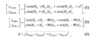
where θ = joint angle and d = distance between the origins of the thumb and index finger MCP joint base frame.
Glove Design
Figure 3 shows the glove prototype. We focused the underlying glove design on fitting a reasonable variety of hand sizes [25]. We created several glove prototypes using various material combinations but determined that spandex (a Lycra and cotton combination) provided maximum flexibility in addition to being lightweight and soft. We
incorporated open-ended fingertips to allow haptic feedback and assist in grasping manipulation and hand shaping. We added a commercially available wrist support (Sammons Preston; Bollingbrook, Illinois) for stability and to ensure reliable positioning of the glove. This splint helps secure the glove at a consistent position with respect to the wrist and prevents unwanted slipping during active usage. To measure the joint angles needed for the grasp prediction model, we positioned four bendsensing resistors (Flexpoint Sensor Systems, Inc; Draper, Utah) over the PIP and MCP joints of the thumb and index finger (Figure 3).
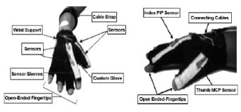
Figure 3. Sensorized glove front and side views. Glove is lightweight and made of cotton and Lycra with four flexible sensors used to measure thumb and index finger joint angles. Sensor sleeves have 30 slots on index finger and 20 slots on thumb. Width of each sleeve is 5 mm with tolerance of ±1 mm. MCP = metacarpophalangeal, PIP = proximal interphalangeal.
We used accuracy and placement of sensors as two key constraints for sensor selection. Accuracy is crucial for properly acquiring data. It was important that the sensors did not interfere with grasping or reduce the glove�s capability to accommodate varying finger lengths. We used bend sensors (Flexpoint Sensor Systems, Inc; Draper, Utah) because they met these needs [26]. These resistive sensors are accurate position sensors with significantly smaller, lighter, and more robust profiles than other possible sensors, such as precision trimming potentiometers (Bourns, Inc; Riverside, California). Bend sensors have a small profile (0.127 mm x 5 mm x varying lengths) that allows them to fit snugly in the glove sleeves without additional mounting. We used customsewn nylon sleeves (5 mm wide) to secure the sensors. The sleeves act as a securing guide and allow the sensors to slide back and forth on top of the fingers as they flex and extend but also prevent unwanted side drifting. Both ends of the sensor sleeves have Velcro that, together with the slots, allow flexible adjustments for varying finger lengths (e.g., the sleeves can be pulled tighter as needed for shorter digits).
We chose FES to facilitate hand opening because of its capability to target specific muscle groups. The features of the FES device, such as low weight, portability, and use of low-profile gelled, self-adhesive electrodes, made it suitable for straightforward integration with the ADLER system. The FES glove system facilitates hand opening through recruitment of the muscles involved during a particular action, for instance, the extensor muscle family for wrist extension and hand opening. These actions reinforce direct involvement of the impaired hand and promote the use of strategies similar to those used before the stroke. Finally, the estimated cost of this device is $49.99, meeting our low-cost goal.
Glove Control Design
We connected the sensors on the glove to a custom circuit controlled by a pair of dedicated ATmega8 RISC (reduced instruction set computer) microprocessors (Atmel Corp; San Jose, California) (Figure 4). We chose the dedicated microprocessor configuration by considering a control scheme where one microprocessor focuses on collecting, storing, and transmitting sensor data and the other focuses on controlling the stimulation amplitude of the FES device. This dual dedicated microprocessor setup is crucial in managing processor resources and reducing potential bottlenecks in memory allocation and processor time, especially for measuring real-time joint angle, predicting grasp aperture, and controlling stimulation assistance. Data transmission between the microprocessors and computer occurs through an RS-232 protocol [27]. A custom manual trigger that controls the simulator allows user-initiated stimulation.
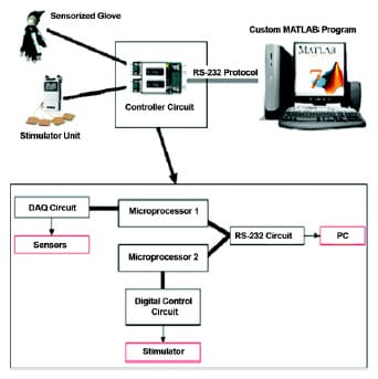
Figure 4. Functional electrical stimulation sensorized glove system with major
components and control flow diagram. Glove data combined with data from
unit are collected with serial communication and processed with use of
custom MATLAB program. Two ATmega8 microprocessors (Atmel Corp; San Jose, California)
mediate collection process. DAQ = data acquisition, PC = personal computer.
The FES device we used with this system is a U.S. Food and Drug Administration-approved dual-channel functional electrical muscle stimulator called the EMS7500 Digital Muscle Stimulator (WisdomKing.com, Inc; Oceanside, California) (Figure 5). This device is handheld, powered through a 9 V battery, and capable of providing a maximum output of 80 mA. The output signal is an asymmetric square pulse with a frequency range between 2 and 120 Hz and a width range of 50 to 300 μs. We modified the FES device to be software-controlled by a personal computer-based custom MATLAB (The Math- Works; Natick, Massachusetts) graphical user interface (GUI) (Figure 5). The GUI consists of two main portions. The first is the controller portion for the FES device and the second is the controller portion for the bend-sensor data collection unit. The device�s settings and amplitudes are software-adjusted with a finer gradation, enabling a quantitative recording of the amount of stimulation administered. The GUI program accepts the incoming data from the microprocessor and sends appropriate controller signals for the device.
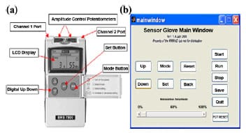
Figure 5. (a) EMS7500 functional electrical muscle stimulator (WisdomKing.com,
Inc; Oceanside, California). (b) Personal computer-based graphical
user interface. LCD = liquid crystal display.
Glove Calibration and Setup
Before the start of the task, we asked the subjects to don the glove and then we calibrated it. Calibration is performed for two postures: first, when the hand is fully open, and second, when the hand is fully closed. For each of these postures, we recorded the joint angles using a goniometer and recorded bend sensor information with the MATLAB program. In addition, we measured the thumb and index finger from the tip of the distal interphalanges to the PIP joint (lt1, li1), from the PIP joint to the MCP joint (lt2, li2), and from the base of the thumb to the base of the index finger.
Glove Validation Procedures
Ten subjects participated in the validation studies. All subjects consented before participating in this study, which was approved by the Institutional Review Board of the Medical College of Wisconsin and Marquette University. Of the 10 subjects, 5 were nondisabled, right-hand dominant males, with a mean age of 39.3 years. The other five
were subjects with stroke (two female and three male), with a mean age of 67.4 years. Two of the subjects with stroke were right-hand impaired and three were left-hand impaired: all had varied levels of muscle weakness and spasticity (Table 1). We clinically assessed the subjects with stroke using the manual muscle test (MMT) and the Modified Ashworth Scale [28�31]. We used the MMT to assess strength and function of select hand and wrist muscles (e.g., forearm supinators, wrist flexors/extensors, and finger flexors/extensors), for which scores range from 0 to 5, with a score of 0 for a subject with no movement or excitation and a score of 5 for a subject able to hold a position against test-applied resistance. We used the Modified Ashworth Scale to measure spasticity for the hand and wrist muscles (e.g., forearm supinators, wrist flexors/extensors, and finger flexors/ extensors). The scores range from 0 to 4, with a score of 0 being flaccid with no spasticity and a score of 4 having the highest level of spasticity.

Table 1. Clinical assessment of manual muscle test (MMT) and Modified Ashworth Scale
scores (AshW) for subjects (S) with stroke.
We validated the sensorized glove system in three trials:
- A static validation that compared the glove with a standard goniometric tool to assess the repeatability and validity of the system.
- A dynamic validation that compared the glove with a standard dynamic measurement tool to assess the capability of the FES glove system to accurately collect joint-angle data and predict an accurate grasp aperture during a real-life activity, such as reaching out for various objects for the five nondisabled subjects.
- A usability validation that examined subjects� hand opening with and without the modified FES device to determine whether the glove�s stimulation component can cause hand opening in the five subjects with stroke who lacked this ability.
For our static and dynamic validation procedures, we attempted to determine not only face validity but also criterion- oriented validity, i.e., the relationship of our measurement instrument to gold standard instruments such as the goniometer and a popular infrared camera-based plus passive markers motion analysis system [32�35].
Static and Dynamic
We instrumented the five nondisabled subjects with the sensorized glove on their nondominant hand and a set of reflective markers based on the Bilateral Upper-Limb Kinematic Model (UL Model) [36]. This model consists of 12 reflective markers placed on key bony anatomical landmarks on the upper limb (Figure 6). We modified the original UL Model by including an additional eight markers that were placed on the tips of the index finger, thumb, and on the fifth and third metacarpals to provide data pertaining to the grasp aperture and hand position in three-dimensional (3-D) space.
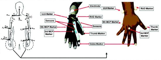
Figure 6. Extension of upper-limb kinematic model with additional markers on third and fifth
metacarpophalangeal (MCP) joints and tips of index finger and thumb. Circle indicates additional
markers used on hand. Directions are based on perspective of viewer.
ec = elbow joint center, mlacr = left acromion process marker, mlelb = left olecranon marker,
mlrad = left radial styloid marker, mracr = right acromion process marker, mrelb = right olecranon marker,
mrrad = right radial styloid marker, mstrn = sternal notch marker, RAD = radial styloid,
Sc = shoulder center, tc = trunk center, ULN = ulnar styloid, xle = x-axis of left elbow,
xlw = x-axis of left wrist, xre = x-axis of right elbow, xrw = x-axis of right wrist,
xt = x-axis of trunk frame, zle = z-axis of left elbow, zlw = z-axis of left wrist,
zre = z-axis of right elbow, zrw = z-axis of right wrist, zt = z-axis of trunk frame,
(x) = circumference of shoulder around acromion and axilla.
Subjects sat at a table with their hands resting on the surface and their elbows at 90� of flexion (Figure 7(a)). We outlined the position of their hands using a permanent marker on the table overlay. To assess repeatability and sensitivity of the glove in a static setting, we collected individual joint-angle measurements using a common clinical handheld 5.5 in. finger goniometer (Sammons Preston; Bollingbrook, Illinois) as our objective measurement device for the individual joint angles. This handheld goniometer is a standard tool used in clinical environments to measure joint angles of the digits. The accuracy of this device is up to within 5°. To assess dynamic repeatability and accuracy, we used a 15-camera VICON motion analysis system (VICON; Los Angeles, California) as the validated objective measurement tool for grasp-aperture data collection [20�21,33,36]. We then compared data from these two systems with the data from the sensorized glove and grasp-prediction model. We collected data from the VICON system at a rate of 120 Hz, and the glove system collected data at a rate of 28 Hz.
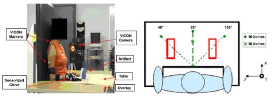
Figure 7. Setup for validation tests for dynamic and static portions.
Subject is instrumented with VICON markers (VICON; Los Angeles, California) on
arm and hand and seated at table. Glove is worn on right hand for illustration purposes only.
For static assessment, we collected data while the five nondisabled subjects maintained their hands fully open and closed. These postures are typically used to measure finger range of motion [34�35]. For each of the hand postures, we collected measurements three times and for 5 seconds for each trial using the glove and goniometer. For the two hand postures, we did not coach the subjects or ask them to have their hands at any pa
rticular fixed position, but rather we asked them to open and close their hands at comfortable postures. For the handopen postures, the subjects placed the palm of their hand on the table and kept it as flat as possible. For the dynamic portion of the study, we presented the subjects with a series of four tasks (Figure 8). The first three tasks consisted of reaching out and picking up objects, and the last task consisted of performing a drinking sequence. The artifacts used in the first three tasks consisted of a pen, ball, and vertical cylinder that we placed at three different orientations of 45°, 90°, and 135° from the subject�s midline (Figure 7(b)). We placed these objects 16 in. from the edge of the table at the midline in a given orientation. During the “pickup” tasks, the subject reached out to the object, picked it up, returned it, and returned to the starting position. We placed more emphasis on the geometric properties of the artifacts than on their functionality. We chose these three shapes because they represented items used frequently during ADLs in the home setting. For the drink task, the subject reached out to pick up a cup located 10 in. from the edge of the table, took a sip, returned the cup, and then returned to rest. This task evaluated our glove within an actual functional task. The subjects repeated all of the tasks three times.

Figure 8. Task event details.
Usability
The last part of the validation study consisted of a usability analysis study with the five subjects with stroke. The study helped qualify the capability of the FES component of the glove to facilitate hand opening and the capability of the modified FES device to provide a quantitative stimulation level administered for each patient. We obtained subjects� spasticity and strength scores, assessed by a licensed physiatrist using the MMT and the Modified Ashworth Scale score, respectively (Table 1), to examine the relationship between these measures and the stimulation amplitude we applied to facilitate grasp aperture.
For hand opening, we located the extensor digitorum muscle group on the forearm by passive activation of the fingers [35]. We placed a pair of stimulator electrodes at the extensor muscle belly [31,34]. We then turned on the FES device and set its amplitude to 0. We did this to ensure that neither the subject nor the experimenter was accidentally shocked or hurt during setup. We also instrumented the subject with the reflective markers and sensorized glove worn over the nondominant hand. Once setup was complete, we connected the electrodes to the FES device. We informed the subject that the amplitude would gradually increase until a comfortable threshold that opened the hand with minimal pain was determined. We then gradually increased the administered stimulation at the predefined step of 0.8 percent, or 640 μA. At every increment, we asked the subject if the level was bearable. Once the subject obtained a comfortable stimulation threshold, we recorded the amplitude percentage. We used the percentage instead of the raw current to compare opening strength across subjects.
We assessed three hand postures. For the first posture, the subjects rested their hands in the start position on top of the table. This start position consisted of the subject placing his or her hand on the table and relaxing without eliciting any muscle contractions of the hand. The second posture required subjects to open their hands to the best of their ability. The final posture required subjects to open their hands to the best of their ability with FES.
Validation Data Analysis
We analyzed the data for the static and dynamic portions of the validation study using MATLAB, Microsoft Excel (Microsoft Corp; Redmond, Washington), and StatView (SAS Institute, Inc; Cary, North Carolina). We processed data using custom-written programs in MATLAB, Microsoft Excel, and VICON. We reconstructed the kinematic data for position and orientation of the trunk, shoulder, elbow, and wrist using 3-D coordinates in space together with the UL Model and extended markers. We filtered the VICON data (size 20 Woltering filter) [33�34,36�38]. The Woltering filter was designed specifically for kinematic gait analysis. It is equivalent to a double Butterworth filter but can process data sets of unequal sampling intervals. We transformed the raw glove data into appropriate joint angles, which we then implemented through the proposed grasp model to obtain the grasp-aperture information. We used a digital zero phase four-point averaging filter to process the glove data.
To investigate the correlation between the joint angles for the hand-open and -closed postures for the static study, we obtained the mean and standard deviation (SD) for each subject in each hand position. We compared the data from the glove with those from the handheld goniometer for each of the three trials for the individual joint angles. The SD for all the data measured from the handheld goniometer was 0 because of its accuracy of only 5°. We divided the data set into four main categories: finger type (index and thumb), joint types (PIP and MCP), device type (glove and goniometer), and grasp posture (open and closed). We compared all three trials for each of the data sets using the repeated measures analysis of variance (ANOVA) followed by Fisher�s PLSD (protected least significant difference) as a post hoc test for all five nondisabled subjects using a significance value of 0.05 [37]. The data set met the normality conditions for the ANOVA.
To assess the correlation between the glove data with the VICON data for the grasp aperture during hand task performance, we compared these data to determine whether a significant difference existed. The analysis of the functional tasks required each data set to be segmented according to the timing of the respective events in each task (Table 1). We calculated the patient average for each of these segments across each trial. Next, we performed a repeated measures ANOVA with p = 0.05 to determine the correlation between the grasp aperture and the measurement devices, across subjects and across events. We used the Scheff� test as a post hoc analysis for the dynamic tasks [37]. The data set for the nondisabled subjects met the normality conditions for the ANOVA.
We collected the usability portion of the analysis for the glove data by comparing the grasp aperture in the nonstimulated posture with the stimulated posture. We performed a repeated measures ANOVA using a significance value of 0.05; similarly, we calculated the Scheff� test for post hoc analysis. The data set met the normal conditions for the ANOVA.
Validation ResultsZ
Static
We tested the null hypothesis that no difference existed between the glove data measurement of finger joint angles and the handheld goniometer data for each finger and finger joint. The results for the static validation study for the thumb and index finger joints in the open and closed postures are shown in Figure 9 and Tables 2 and 3. In examining Table 3, we saw that from the ANOVA calculations, a significant difference existed across the finger types (p = 0.002), joint types (p = 0.007), and grasp posture (p < 0.001). In examining the data for the device type, we noticed that no significant difference existed between the glove and goniometer data (p = 0.93).
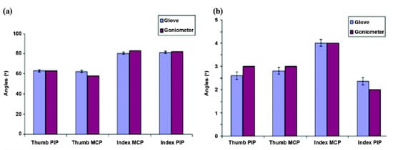
Figure 9. Aver
age joint angles for (a) hand-closed posture and (b) hand-open posture.
Error bars represent standard error. MCP = metacarpophalangeal, PIP = proximal interphalangeal.
In Table 2, we show that for the closed posture, subject 4 had the lowest measured joint angle for the thumb PIP at 30°. Subject 4 also had the highest joint angle of 101.24° for the index PIP joint. The table also indicates that during the hand-open posture, even though all subjects had their hands fully flat on the table to the best of their abilities, not all joint angles were at 0°. Subject 4 had the highest joint angle of 6.48° for the thumb MCP joint. In examining the data for the static validation study, we observed that the results obtained from the glove were similar to those obtained from the handheld goniometer. The measurements for subject 5�s index PIP joint had the largest SD of 2.78°. In looking at the overall results, we observed that the measured data from the glove and goniometer correlated closely.

Table 2. Comparison of joint angles (mean � standard deviation) measured from
sensorized glove (GL) and handheld goniometer (GO) for five nondisabled subjects.

Table 3. Fisher�s PLSD (protected least significant difference) for finger type, joint type,
grasp posture, and device type using significance value of 0.05 for five nondisabled subjects.
Critical difference is 2.782.
Dynamic
We tested the null hypothesis that no difference existed between the predicted grasp aperture from the glove data and from the VICON data for each task in a given direction. Figure 10 shows an example result of the drink task for the glove versus the motion analysis data. Figures 11 and 12 show us the mean ± SD data for the five subjects for grasping and lifting the ball, cylinder, and pen located at 45°, 90°, and 135° by events and for completing the functional drink task by events. We observed both motion analysis and glove data consistent across the three angles, suggesting repeatable measurements. We observed the largest aperture in the performance of the cylinder task and the smallest apertures during the pen task. The largest differences exist for the cylinder task, where we predicted consistently greater glove grasp aperture than that predicted by the motion analysis for all events. Significant differences also existed for the pen task at 45° and 135°, where the grasp aperture data predicted for events 2 and 3 (grasp and lift of the object) were consistently lower than the motion analysis data. We also saw this difference on the same two events for the drink task.
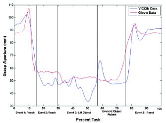
Figure 10. Example plot of grasp aperture collected by glove versus VICON
(VICON; Los Angeles, California) grasp aperture. Grasp-aperture prediction graph shows
how once object is grasped in event 2, prediction should remain static until object is released.
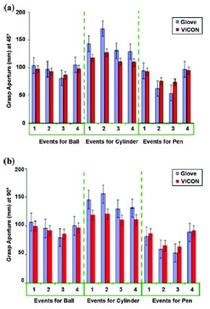
Figure 11. Grasp aperture for objects located at (a) 45° and (b) 90° for both glove
and VICON (VICON; Los Angeles, California) data for ball, cylinder, and pen. Grasp aperture
for pen was consistently less than predicted apertures for all other objects.
Differences between glove and VICON prediction of grasp aperture were greater
for all events of cylinder object. Events 1�4: reach for object, grasp/lift object,
return object, and return to rest, respectively. Error bars represent standard error.
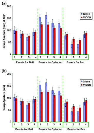
Figure 12. (a) Grasp aperture for objects located at 135° for both glove and VICON (VICON; Los Angeles California) data for ball, cylinder, and pen. Patterns seen at 135° are similar to patterns seen at 45° and 90° in Figure 11. Events 1�4: reach for object, grasp/lift object, return object, and return to rest, respectively. (b) Grasp aperture for functional drink task. Differences between grasp aperture predicted by glove were smaller than VICON prediction for 2 of 5 events, namely events 2 (grasp cup) and 3 (bring cup to mouth). Events 1�5: reach for cup, grasp cup, lift cup to mouth, replace cup, and return to rest, respectively. Error bars represent standard error.
Table 4, the calculated ANOVA and post hoc results of the dynamic validation study, shows a significant difference between events (p < 0.001) and tasks (p < 0.001), but no significant differences exist between devices (p = 0.16) and subjects (p = 0.65). The results from our post hoc analysis (using a significance level of 5%) support our ANOVA findings, in that differences exist between events and tasks but not devices and subjects.

Table 4. Analysis of variance and post hoc results for dynamic validation study
for five nondisabled subjects.
Usability
We hypothesized that the stimulation component of the glove could provide the subjects with stroke with adequate hand opening. We further hypothesized that the modified version of the FES device could provide a quantitative level of stimulation administered for each subject. We tested the null hypothesis that subjects who could not complete the tasks without assistance could have a similar grasp aperture with and without stimulation. Table 5 shows the average results of the stimulation trials. We can see that, overall, the assisted hand-open postures had si
gnificantly larger grasp apertures than nonassisted hand-open postures. Comparing the assisted and nonassisted postures of hand open using ANOVA showed significant differences, with p = 0.001.

Table 5. Grasp aperture (cm, mean � standard deviation) for each of three trials
for five subjects with stroke for three hand postures.
Discussion
We presented the model and prototype of an FES grasp glove that could be used as a tool for measuring hand opening and closing during functional tasks inside and outside of a robotic therapy environment for stroke rehabilitation. We combined the glove with a modified muscle stimulator to provide assistance for hand opening during functional grasping. We conducted two trials designed to validate the system to determine whether its accuracy equaled standard tools during static and dynamic validation studies. We also conducted a trial to verify whether the FES assistive portion of the system was usable by subjects with stroke and able to open spastic and nonspastic limbs. Overall, our results indicated that the grasp aperture measurement for hand posture was just as accurate as static and dynamic standard measurement tools.
For static validation, a significant difference did not exist between the predicted glove grasp-aperture data and the data measured using the goniometer tool, which suggests that the glove�s predicted grasp-aperture measurement for hand posture was just as accurate as this standard tool. Significant differences we saw for the finger and joint types show that these joints have different angles that contribute to the overall grasp aperture achieved. We expected differences across the grasp types, since a larger difference existed between the joint angles when the hand was fully open versus when it was fully closed. The study also showed us that the data signals obtained from the system were stable over time and that little or no noise factor influence was found. This finding is crucial in determining the stability of the signal because it influences the future accuracy, especially in the dynamic validation portions of the task where potential errors caused by noise interference could potentially be hard to detect.
Although the glove was stable in a static environment, we anticipated that the dynamic environment, i.e., the subject performing tasks with the glove, would challenge stability of the sensors and the predictive capability of the model because of movement of the sensors [33]. Therefore, testing the glove dynamically was important. This observation also suggests that we could improve the static validation protocol by having the subject remove the glove and don it again to determine its impact on the model�s predictive capability.
For the dynamic validation part of the study, no significant differences existed between the predicted glove grasp-aperture data and the data predicted by the infrared video camera motion analysis process. This finding suggests that the glove�s predicted grasp-aperture measurement for hand posture was just as accurate as this standard tool. We did observe significant differences amongst events and tasks. We expected these differences because the grasp aperture is largest during preshaping and varies during manipulation of the object, often depending on the object size [20]. The differences between events showed us that an influence of object type on the aperture size exists. We can also see this difference in the third calculation; for objects that were larger (cylinder), we used a larger grasp aperture, while for objects that were smaller (pen), we used a significantly different and smaller aperture. Since our statistical analysis indicated no significant differences between subjects, we conclude that observed differences were due to chance and each subject initiated the same strategy during prehension for each of the tasks.
The glove tended to predict higher grasp aperture for the cylinder task and lower grasp apertures for grasp and lift events for the grasping of a pen and a cup handle (Figures 11 and 12). One possible reason was that the distance measured from the VICON system was based on the distance dictated by the center of the reflective markers and their locations. The marker positions were not on the tips of the fingers, but rather on top of the fingernails. This position reduced the total length of each digit compared with the grasp aperture that we obtained from the glove and the model where the grasp aperture is predicted as the distance between the tips of the thumb and index finger. In addition, marker size added to the difference, because it influenced the distance of the marker�s center position relative to the tip of the finger. Another possible source of these differences could have been sensor drift or slipping. Although we minimized sensor drift and slipping by creating and implementing the special sleeves that fit the sensors snugly, additional drifts could have arisen in the functional setting, in that the glove expanded and contracted during reaching and manipulation events. Another possible source of these grasp prediction differences could have been marker dropout. Marker dropout is a common problem in motion analysis [33,36] and occurs when the reflective markers are hidden from the cameras. For our tasks, the reflective markers tended to drop out during initial grasp and manipulation of the object, where a higher probability existed that the object itself might block the markers. We compensated for marker dropout during the VICON postprocessing through an interpolation process. If marker dropout is large, the interpolation will introduce larger errors. We did observe that marker dropout was more significant for grasping events than for other events. Therefore, we expected the glove to predict grasping events better. Further dynamic validation of the glove is needed and will be completed in the near future. Examining the glove�s accuracy is important in measuring graspaperture change over time as is the capability of the sensor system to continue to accurately predict grasp aperture after long-term use.
The results obtained from our FES validation study showed that the system did indeed have an improved effect in facilitating hand opening. We clearly saw this in the hand open with assistance postures, where, with assistance from the device, hand opening was achieved and the contrary was not possible. This finding helps to reaffirm our proposed hypothesis and show that the FES portion of our glove is indeed capable of facilitating hand opening. We anticipate some subjects with severe spasticity may not benefit from this assistance. The range of subjects with stroke that can be assisted and the range of grasping tasks that can be supported by FES need to be examined.
The combined results of these studies indicate that the developed system can be integrated into the ADLER system for reaching and grasping tasks during stroke rehabilitation. Additional studies are needed to further evaluate the system during task-oriented therapy.
Conclusions
This article examines the design, development, and validation of a custom-made sensorized glove system and its custom grasp prediction model. The validation results show that this system was accurate, stable over time, and repeatable. In addition, the validation studies also helped show the capability of this glove system for real-time tracking and the model for predicting grasp aperture. The FES validation portion proved that the system can deliver qualitative FES to the subject and that this does indeed help with hand opening. The efficacy of the system is
crucial, in that the system serves as a tracking tool that can provide not only real-time functional grasp assistance but also performance tracking for the ADLER system. The next phase will be full integration with our robotic therapy device.
Acknowledgments
Author Contributions:
Study concept and design: D. E. Nathan, M. J. Johnson. Acquisition, analysis, and interpretation of data: D. E. Nathan, M. J. Johnson.
Clinical interactions with subjects: D. E. Nathan, M. J. Johnson, J. R. McGuire.
Glove development: D. E. Nathan, M. J. Johnson, J. R. McGuire.
Drafting of manuscript: D. E. Nathan, M. J. Johnson, J. R. McGuire.
Critical revision of manuscript for important intellectual content: D. E. Nathan, M. J. Johnson, J. R. McGuire.
Study supervision: D. E. Nathan, M. J. Johnson.
Financial Disclosures: The authors have declared that no competing interests exist. No author had any paid consultancy or any other conflict of interest with this article.
Funding/Support: This material was based on work supported by the National Institutes of Health (grant 2203792) through the Rehabilitation Institute of Chicago, the Medical College of Wisconsin Research Affairs Committee (grant 3303017), and Advancing a Healthier Wisconsin (grant 5520015) for the ADLER project.
Additional Contributions: We thank John Anderson, Kimberly Wisneski, and Greg Millar for their work leading to the development of the ADLER environment; Dr. Gerald Harris, Dr. Brooke Hingtgen, and Dr. Jason Long for assisting us in the Orthopaedic and Rehabilitation Engineering Center (OREC) Human Motion Analysis Laboratory, Milwaukee, Wisconsin; Dr. Guennady Tchekanov for assisting with data collection; and the Clement J. Zablocki Department of Veterans Affairs Medical Center, Milwaukee, Wisconsin, for their infrastructure support of the Rehabilitation Robotic Research Design Laboratory, which is also supported by the Medical College of Wisconsin, Milwaukee, Wisconsin, and Marquette University, Milwaukee, Wisconsin.
Portions of this article were published and presented in Nathan DE, Johnson MJ. Design and development of a grasp assistive glove for ADL-focused robotic assisted therapy after stroke. Proceedings of the 10th IEEE International Conference on Rehabilitation Robotics; 2007 Jun 18�22; Noordwijk, the Netherlands.
Participant Follow-Up: The authors plan to inform participants of the publication of this study.
References
- Thom T, Haase N, Rosamond W, Howard VJ, Rumsfeld J, Manolio T, Zeng ZJ, Flegal K, O�Donnell C, Kittner S, Lloyd-Jones D, Goff DC Jr, Hong Y, Adams R, Friday G, Furie K, Gorelick P, Kissela B, Marler J, Meigs J, Roger V, Sidney S, Sorlie P, Steinberger J, Wasserthiel-Smoller S, Wilson M, Wolf P; American Hearth Association Statistics Committee and Stroke Statistics Committe. Heart disease and stroke statistics�2006 update: A report from the American Heart Association Statistics Committee and Stroke Statistics Committee. Circulation. 2006;113(6):e85�151. [PMID: 16407573] Errata in: Circulation. 2006;113(14): e696. Circulation. 2006;114(23):e630.
- Bach-y-Rita P. Theoretical and practical considerations in the restoration of function after stroke. Top Stroke Rehabil. 1999;8(3):1�15. [PMID: 14523734]
- Liepert J, Bauder H, Wolfgang HR, Miltner WH, Taub E, Weiller C. Treatment-induced cortical reorganization after stroke in humans. Stroke. 2002;31(6):1210�16. [PMID: 10835434]
- Nudo RJ. Functional and structural plasticity in motor cortex: Implications for stroke recovery. Phys Med Rehabil Clin N Am. 2003;14(1 Suppl):S57�76. [PMID: 12625638] DOI:10.1016/S1047-9651(02)00054-2
- Cramer SC, Nelles G, Schaechter JD, Kaplan JD, Finklestein SP, Rosen BR. A functional MRI study of three motor tasks in the evaluation of stroke recovery. Neurorehabil Neural Repair. 2001;15(1):1�8. [PMID: 11527274] DOI:10.1177/154596830101500101
- Krebs HI, Volpe BT, Ferraro M, Fasoli S, Palazzolo J, Rohrer B, Edelstein L, Hogan N. Robot-aided neurorehabilitation: From evidence-based to science-based rehabilitation. Top Stroke Rehabil. 2002;8(4):54�70. [PMID: 14523730] DOI:10.1310/6177-QDJJ-56DU-0NW0
- Lum PS, Burgar CG, Shor PC. Evidence for improved muscle activation patterns after retraining of reaching movements with the MIME robotic system in subjects with poststroke hemiparesis. IEEE Trans Neural Syst Rehabil Eng. 2004;12(2):186�94. [PMID: 15218933] DOI:10.1109/TNSRE.2004.827225
- Loureiro R, Amirabdollahian F, Topping M, Driessen B, Harwin W. Upper limb robot mediated stroke therapy� GENTLE/s approach. J Auton Robots. 2003;15(1):35�51. DOI:10.1023/A:1024436732030
- Patton JL, Dawe G, Scharver C, Mussa-Ivaldi FA, Kenyon R. Robotics and virtual reality: The development of a lifesized 3-D system for the rehabilitation of motor function. Proceedings of the 26th Annual IEEE Engineering in Medicine and Biology Society Conference; 2004 Sep 1�4; San Francisco, CA. Los Alamitos (CA): IEEE Press; 2004.
- Fazekas G, Horvath M, Troznai T, Toth A. Robot-mediated upper limb physiotherapy for patients with spastic hemiparesis: A preliminary study. J Rehabil Med. 2007;39(7): 580�82. [PMID: 17724559] DOI:10.2340/16501977-0087
- Johnson MJ, Wisneski KJ, Anderson J, Nathan D, Smith RO. Development of ADLER: The Activities of Daily Living Exercise Robot. Proceedings of the 1st IEEE/RASEMBS International Conference on Biomedical Robotics and Biomechatronics; 2006 Feb 20�22; Pisa, Italy. Los Alamitos (CA): IEEE Press; 2006.
- Bouzit M, Burdea G, Popescu G, Boian R. The Rutgers Master II�New design force-feedback glove. IEEE ASME Trans Mechatron. 2002;7(2):256�63. DOI:10.1109/TMECH.2002.1011262
- Takahashi CD, Der-Yeghiaian L, Le VH, Cramer SC. A robotic device for hand motor therapy after stroke. Proceedings of the 9th IEEE International Conference on Rehabilitation Robotics Conference; 2005 Jun 28�Jul 1; Chicago, IL. Los Alamitos (CA): IEEE Press; 2005.
- Worsnopp TT, Peshkin MA, Colgate JE, Kamper DG. An actuated finger exoskeleton for hand rehabilitation following stroke. Proceedings of the 10th IEEE International Conference on Rehabilitation Robotics; 2007 Jun 13�15; Noordwijk, the Netherlands. Los Alamitos (CA): IEEE Press; 2007.
- Loureiro RC, Harwin WS. Reach and grasp therapy: Design and control of a 9-DOF robotic neuro-rehabilitation system. Proceedings of the 10th IEEE International Conference on Rehabilitation Robotics; 2007 Jun 13�15; Noordwijk, the Netherlands. Los Alamitos (CA): IEEE Press; 2007.
- Dovat L, Lambercy O, Johnson V, Salman B, Wong S, Gassert R, Burdet E, Leong TC, Milner T. A cable driven robotic system to train finger function after stroke. Proceedings of the 10th IEEE International Conference on Rehabilitation Robotics; 2007 Jun 13�15; Noordwijk, the Netherlands. Los Alamitos (CA): IEEE Press; 2007.
- Ulrich K, Eppinger SD. Product design and development. 3rd ed. Philadelphia (PA): McGraw-Hill; 2003.
- Cutcosky MM. On grasp choice, grasp models, and the design of hands for manufacturing tasks. IEEE Trans Rob Autom. 1989;5(3):269�79. DOI:10.1109/70.34763
- Mason CR, Gomez JE, Ebner TJ. Hand synergies during reach-to-grasp. J Neurophysiol. 2001;86(6):2896�2910. [PMID: 11731546]
- Gentilucci M. Object familiarity affects finger shaping during grasping fruit stalks. Exp Brain Res. 2003;149(3): 395�400. [PMID: 12632242]
- Gentilucci M, Caselli L, Secchi C. Finger control in the tripod grasp. Exp Brain Res. 2003;149(3):351�60. [PMID: 12632237]
- Kurillo G, Bajd T, Kamnik R. Static analysis of nippers pinch. Neuromodulation. 2003;6(3):166�75. DOI:10.1046/j.1525-1403.2003.03026.x
- Craig JJ. Introduction to robotics: Mechanics and control. 3rd ed. Upper Saddle
River (NJ): Pearson/Prentice Hall; 2005. - Wu G, Van Der Helm FC, Veeger HE, Makhsous M, Van Roy P, Anglin C, Nagels J, Karduna AR, McQuade K, Wang X, Werner FW, Buchholz B; International Society of Biomechanics. ISB recommendation on definitions of joint coordinate systems of various joints for the reporting of human joint motion�Part II: Shoulder, elbow, wrist and hand. J Biomech. 2005;38(5):981�92. [PMID: 15844264] DOI:10.1016/j.jbiomech.2004.05.042
- Tilley AR. The measure of man and woman: Human factors in design. Rev. ed. Chichester (UK): Wiley; 2002.
- Simone LK, Kamper DG. Design considerations for a wearable monitor to measure finger posture. J Neuroeng Rehabil. 200;2(1):5. [PMID: 15740622]
- Nelson M. Serial communications developer�s guide. 2nd ed. Foster City (CA): IDG Books Worldwide; 2000.
- Radomski MV, Trombly Latham CA. Occupational therapy for physical dysfunction. 5th ed. London (UK): Lippincott Williams & Wilkins; 2001.
- Braddom R, Peterson AT. Handbook of physical medicine and rehabilitation. Philadelphia (PA): Saunders; 2004.
- Cuccurullo SJ. Physical medicine and rehabilitation board review. New York (NY): Demos Medical Publishing; 2004.
- O�Young B, Young MA, Stiens SA. Physical medicine and rehabilitation secrets. 2nd ed. Philadelphia (PA): Hanley and Belfus Publishing; 2001.
- Portney LG, Watkins MP. Foundations of clinical research: Applications to practice. 2nd ed. Upper Saddle River (NJ): Prentice Hall; 2000.
- Anglin C, Wyss UP. Review of arm motion analyses. Proc Inst Mech Eng H. 2000;214(5):541�55. [PMID: 11109862] DOI:10.1243/0954411001535570
- Trombly Latham CA. Occupational therapy of physical dysfunction. Baltimore (MD): Williams & Wilkins; 1995.
- Nigg BM, Herzog W. Biomechanics of the musculo-skeletal system. 3rd ed. Hoboken (NJ): John Wiley & Sons; 2007.
- Hingtgen B, McGuire JR, Wang M, Harris GF. An upper extremity kinematic model for evaluation of hemiparetic stroke. J Biomech. 2006;39(4):681�88. [PMID: 16439237] DOI:10.1016/j.jbiomech.2005.01.008
- Gravetter FJ, Wallnau LB. Essentials of statistics for the behavioral sciences. 4th ed. Pacific Grove (CA): Wadsworth/ Thompson Learning; 2002.
- Woltring H. Smoothing and differentiation techniques applied to 3D data. In: Allard P, Stokes IA, Blanchi JP, editors. Three-dimensional analysis of human movement. Champaign (IL): Human Kinetics; 1995.


