Thay Q. Lee, PhD; Bruce Y. Yang, BS; Matthew D. Sandusky, BS; Patrick J. McMahon, MD
Orthopedic Biomechanics Laboratory, Long Beach VA Healthcare System, CA; Department of Orthopedic Surgery,
University of California, Irvine, CA; Department of Orthopaedic Surgery, University of Pittsburgh, Pittsurgh, PA
Abstract
The objective of this study was to determine the effects of tibial rotation on in situ strain in the peripatellar retinaculum and patellofemoral contact pressures and areas. Patellofemoral joint biomechanics demonstrate a strong correlation with the etiology of patellofemoral disorders, such as chondromalacia, and are significantly influenced by tibial rotation. Six human cadaveric knees were used along with a patellofemoral joint testing jig that permits physiological loading of the knee extensor muscles. Patellofemoral contact pressures and areas were measured with a Fuji pressure-sensitive film, and the changes in in situ strain in the peripatellar retinaculum were measured with four differential variable reluctance transducers. Tibial rotation had a significant effect on patellofemoral joint biomechanics. The data showed an inverse relationship between increasing knee flexion angles and the change in patellofemoral contact pressures and in situ strain with tibial rotation. At higher knee flexion angles, the patella is well-seated in the trochlear groove and the function of the peripatellar retinaculum is minimized and less affected by tibial rotations.
Key words: contact area, contact pressure, in situ strain, patellofemoral joint, tibial rotation.
INTRODUCTION
The mechanism of the patellofemoral joint has been studied extensively by determining the quadriceps force with respect to various physiological activities (1–5). In these earlier studies, the patellofemoral joint was assumed to be a frictionless pulley system; this was later found to be an unrealistic representation (3–7). The quadriceps muscle force acts on the patella with a lever arm that originates at the geometric center of the load-transmitting articular surface. The patellar ligament force, however, acts on the patella with a different lever arm that also originates at the center of the articular surface. These two lever arms vary during movement and are thus not always equal (8). Therefore, the quadriceps muscle force and the patellar ligament force are not equal. Instead, the ratio of the two forces varies in a complex manner as a function of knee flexion angle. Below 45º knee flexion, the force in the patellar ligament is greater than in the quadriceps tendon. At knee flexion angles greater than 45º, this phenomenon is reversed (8). These complexities in the patellofemoral joint necessitate anatomically based loading of the patellofemoral joint during in vitro testing (9,10).
Patellofemoral joint mechanics demonstrate a strong correlation with the etiology of patellofemoral disorders, such as chondromalacia (11–13). Chondromalacia of the patella is considered the primary precursor to arthrosis of the knee (14). Chondromalacia of the patella can result from any condition of the patella in which the normal physiology of the quadriceps mechanism is disturbed, where abnormal stress distribution on the articulating surfaces of the patellofemoral joint occurs. These aberrations in the patellofemoral joint force system can ultimately lead to painful, degenerative joint disease. In addition, imbalance of the patellofemoral mediolateral force, often in conjunction with abnormalities in the bony anatomy of the joint, has also been associated with subluxation of the patella, leading to chondromalacia and arthrosis (6,12,15,16). Furthermore, literature has documented patellofemoral joint disorders resulting from problems associated with soft tissues, the femur and/or the tibia (17–19). However, no studies have quantified the relationship between patellofemoral joint contact pressures and areas and in situ strain in the peripatellar retinaculum, with respect to tibial rotation. The objective of this study was to determine the effects of tibial rotation on in situ strain in the peripatellar retinaculum and patellofemoral contact areas and contact pressures. The null hypothesis is that tibial rotation will have no effect on in situ strain and patellofemoral contact pressures and areas.
METHODS
Specimen Preparation
Six fresh-frozen unmatched cadaveric knees were used in this study. All knees were macroscopically intact with radiographically normal bone structure and no history of knee surgery. The ages of the specimens were estimated to be between 60 and 80 years. The specimens were dissected carefully, removing the skin and subcutaneous tissue and leaving the patellofemoral joint, tibiofemoral joint, and knee capsule intact. Individual components of the extensor mechanism (Vastis Medialis, Vastis Lateralis, Vastis Intermedius, Rectus Femoris, Iliotibial Band) were left intact and clamped to allow application of muscle forces. The superior part of the synovial pouch was liberated to allow placement of Fuji pressure-sensitive film (Fuji Photo Film Co, Ltd., Tokyo, Japan). Specimens were kept moist throughout testing with 0.9-percent saline.
Patellofemoral Joint Testing Jig and Specimen Mounting
The specimen was mounted at full extension, in a custom testing jig (Figure 1). This jig was designed around the Instron materials-testing machine frame (Model #1122, Instron Corp., Canton, MA), which was used to flex the knee and provide the mechanism for anatomical muscle pull (10). The custom testing jig allowed the individual application of extensor muscle forces along the orientation of principle muscle fibers. The tibia and femur of each specimen were placed in mounting cylinders and held in place with fixation pins and diaphyseal bolts. The femur was mounted with its coronal plane parallel to the cylinder and in anatomic valgus
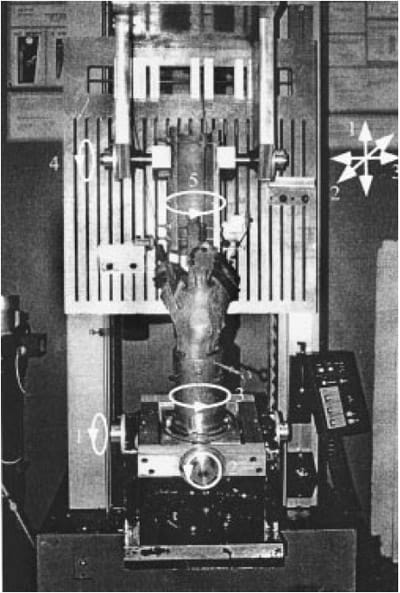
Figure 1. A specimen mounted at full extension in the custom patellofemoral joint testing jig. Numbers indicate degrees of freedom at both the femur and tibia.
Physiological knee motion requires six degrees of freedom at the knee joint. The knee testing jig used in this study permitted five degrees of freedom at the femur (three translational and two rotational) and three degrees of freedom at the tibia (rotational), which permit six degrees of freedom at the knee joint. These degrees of freedom are essential for achieving proper physiological knee flexion. The experimental setup provide
d control over various knee flexion angles and tibial rotations. Knee flexions were confirmed with a goniometer. This experiment was performed at knee flexion angles of 0º, 30º, 60º, and 90º. At each knee flexion angle, measurements of corresponding patellofemoral contact pressures and areas were taken at neutral, 10º and 15º of internal and external tibial rotation. Strain was tracked at increments of 5º up to 20º of internal and external rotation. The differences in the tibial rotation for the contact pressure measurements and strain measurements were necessitated due to the unacceptable artifacts created on the Fuji film at 20º of tibial rotation.
Reference Neutral
Due to the geometry within the knee, the anatomically neutral position of the patella can display a high degree of variability. Therefore, it was essential to establish a protocol that allowed for consistent determination of a “reference” neutral position from specimen to specimen. First, it was necessary to establish the maximal degree of rotation at the tibia within each specimen. The amount of torque used to establish this range was 15 N-m and was applied to each specimen with a torque wrench. The neutral was then determined through the bisection of this maximal range of tibial rotation. This neutral position for each specimen was used as the reference for both change in strains and contact pressures.
Patellofemoral Contact Pressures and Areas
The measurement of contact pressures was accomplished with “Fuji Pre-scale (super-low) pressure-sensitive film” (Fuji Photo Film Co, Ltd., Tokyo, Japan). The film was cut to 5 cm35 cm. The Fuji film comprises two parts, a “transfer sheet” and a “developer sheet.” Each of the two sheets is approximately 0.10 mm in thickness. The sheets of Fuji film were wrapped within a polyethylene bag to protect the film from fluid. The total thickness of the pressure sensitive film in the polyethylene bag was approximately 0.25 mm. This has a negligible effect on the measurement of patellofemoral contact pressures and areas (20). The transfer sheet contains a microcapsule layer, which contains a translucent colorforming substance. When pressure is exerted, the microcapsules break and release their substance into contact with the developer sheet. The density or shade of the color is proportional to the amount of pressure experienced between the two sheets. Fuji’s (super-low) pressure- sensitive film has a range of 0.33–5.41 MPa, as determined in our laboratory. Due to the low-end threshold of 0.33 MPa for the detection of pressure by Fuji film, the contact areas measured are underestimated. There is a 1-percent degree of accuracy when an optical measuring system is used.
AHewlett Packard (Palo Alto, CA) ScanJet IIc color scanner with an optical resolution of 400 dpi was used to image the Fuji film. After the image was scanned, National Institutes of Health (Bethesda, MD) Image Version 1.52 and a Macintosh Power PC were used to digitize and quantify the contact pressures and areas. Each portion of the contact pressure was then processed and saved as a pixel based on a 256-level grayscale range. With the use of the calibration curve provided by the manufacturer, each pixel within the scan was assigned a pressure value. The overall accuracy of Fuji film for measuring contact pressure is within 10 percent.
At each knee flexion angle, measurements for contact pressures were made at the specified “reference” neutral, 10º and 15º of internal and external rotation. Neutral measurements were made by placing the knee in its determined neutral position, placing the Fuji film within the joint, and loading the muscles. However, for the measurements with the tibia in a rotated state, the tibia starts off in the neutral position at the time of Fuji film placement and is then rotated to the specified 10º or 15º. The clamp and pulley system was adjusted at each knee flexion angle to maintain proper anatomic pull. Fuji film was inserted through the superior aspect of the synovial pouch just prior to application of the extensor muscles and was left in for 2 min to allow the film to completely saturate.
All muscles were loaded simultaneously in order to avoid error associated with the irreversible nature of the film. The total extensor muscle loading force was 276 N, with the distribution of forces being based on muscle cross-sectional areas as reported by Wickiewicz et al. (21). The calculated muscle forces were Vastis Medialis, 67 N; Vastis Lateralis, 98 N; Vastis Inter-medius combined with the Rectus Femoris, 111 N. An additional load of 27 N was placed on the Iliotibial Band. All measurements were repeated for verification of the patellofemoral contact patterns. Any film with an indication of a wrinkling artifact was discarded and the measurement repeated.
Strain Measurement
The change in strain was measured with a Differential Variable Reluctance Transducer (DVRT) (Microstrain, Burlington, VT, USA). The DVRT was set in the peripatellar retinaculum with the use of barbs, which were 3 mm in length. Movement between the two barbs was tracked with the use of free-sliding transducer cores. Motion between the cores was registered as a value displayed as a voltage. These voltages were then converted to distances with a calibration curve. The range of motion that the DVRT can accurately track is approximately 0–4 mm. The repeatability of the DVRT is ±1 m, while resolution is 1.5 mm and accuracy is 0.5 percent. The sensitivity of the DVRT is approximately 2 V/mm.
Four DVRTs were used. First, the center of the patella was determined. The DVRT placement was made by placing two DVRTs at positions 1 cm superior from the center, one in the medial portion (Medial Superior) of the retinaculum and one in the lateral portion (Lateral Superior). Two more DVRTs were placed, in a similar fashion, 1 cm inferior from the center of the patella (Medial Inferior, Lateral Inferior). All four DVRTs were placed parallel to the floor, while the knee was at 30º of knee flexion (Figure 2). Analysis of variance (ANOVA) was used for statistical analysis.
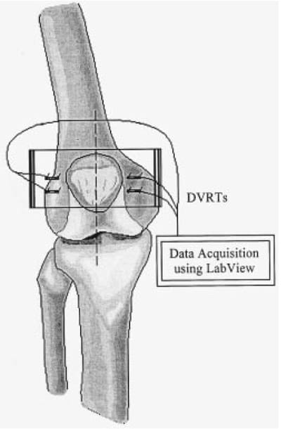
Figure 2. Diagram showing DVRT placement and data acquisition. Diagram is not to scale.
RESULTS
Total Contact Area
Typical patellofemoral contact pressures obtained at 30º KFA for tibial rotations are shown in Figure 3. The range of patellofemoral joint contact area at 0º, 30º, 60º, and 90º flexion was 94.2–158.1 mm2, 174.1–218.8 mm2, 175.6–216.5 mm2, and 147.4–176.6 mm2, respectively. It is important to note that all contact-area measurements are underestimations of the actual articular cartilage contact area, due to limitations within the film to detect pressures below 0.33 MPa.

Figure 3. Typical tibial rotation contact pressures taken at 30º KFA.
Percent change of contact area with respect to tibial rotation is shown in Figure 4. This was determined as the change in area at the externally and internally rotated positions of the tibia, with respect to its corresponding neutral. There was a greater change at 15º of external tibial rotation for 0º, 30º, and 60º of knee flexion, with respect to the corresponding neutral for each knee flexion. The contact area also decreased with increasing knee flexion at 15º of external tibial rotation. At 10º and 15º of internal tibial rotation, there was very little change in contact area with respect to neutral, excep
t at 60º and 90º.
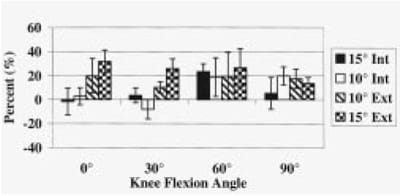
Figure 4. A histogram showing percent change of total contact area with respect to tibial rotation.
Total Contact Pressure
The range of patellofemoral total contact pressures, with respect to tibial rotation, was 0.81–1.23 MPa, 1.04–1.49 MPa, 0.95–1.20 MPa, and 1.01–1.32 MPa, at 0º, 30º, 60º, and 90º, respectively. The total contact pressures were relatively similar at all knee flexions except 0º, where they were the lowest. At each knee flexion, 15º of external tibial rotation had the highest pressure.
Percent change of total contact pressure was determined as change in area at the externally and internally rotated position of the tibia with respect to its corresponding neutral. Percent changes for 10º and 15º of external tibial rotation were inversely proportional to the degree of knee flexion (Figure 5). There were very little changes in pressure for internal rotations of the tibia at any angle of knee flexion.
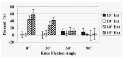
Figure 5. A histogram showing percent change of total contact pressure with respect to tibial rotation.
Top Ten-Percent Peak Pressure The top 10-percent peak pressure represents the highest pressure values corresponding to 10 percent of the total contact area. The range of peak pressures for the top 10-percent contact pressures, with respect to tibial rotation, was 1.45–2.63 MPa, 2.17–3.26 MPa, 1.95–2.77 MPa, and 2.02–3.00 MPa, at 0º, 30º, 60º, and 90º, respectively. The trends seen for top 10-percent peak pressures were essentially identical to those of total pressures. Total contact pressures were relatively similar at all knee flexions except 0º, where they were the lowest. At each knee flexion, 15º of external tibial rotation had the highest pressure.
Again, the percent change of top 10-percent peak pressure was determined as change in the top 10-percent pressure at the externally and internally rotated position of the tibia with respect to its corresponding neutral. Percent changes for 10º and 15º of external tibial rotation were inversely proportional to the degree of knee flexion (Figure 6). There were very little percent changes for internal tibial rotation at any of the knee flexions.
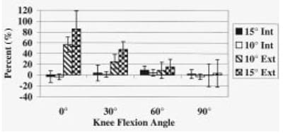
Figure 6. A histogram showing percent change of the top 10-percent peak pressure with respect to tibial rotation.
Changes in In Situ Strain
Change in peripatellar retinacular strain at 20º of internal and external rotation of the tibia exhibited a range of 24.3 to 3.1 percent. With internal rotation of the tibia, strain decreased in the lateral retinaculum; with external rotation, strain decreased in the medial retinaculum with one exception at 0º flexion, the medial inferior DVRT (Table 1).

Table 1. Change in strain of peripatellar retinaculum at 20º of external and internal tibial rotation with respect to neutral tibial rotation (%).
As shown at 30º knee flexion, rotation in a particular direction resulted in decrease of strain on the contralateral side, as previously stated (Figure 7). For example, external rotation of the tibia caused a decrease in strain at the medial superior and medial inferior DVRTs. Internal rotation of the tibia resulted in a decrease in strain at the lateral superior and lateral inferior DVRTs.
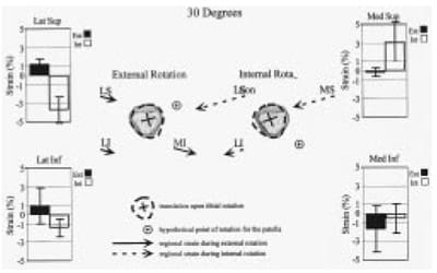
Figure 7. Histograms and illustration showing strain in peripatellar retinaculum with respect to tibial rotation at 30º knee flexion angle. Patella translation upon internal and external rotation is shown through dashed lines that represent the position of patella post tibial translation. Strain within retinaculum is demonstrated through solid and dashed arrows that represent magnitude and direction of strain distribution within four regions of peripatellar retinaculum tested.
DISCUSSION
Tibial rotation had a significant effect on the patellofemoral contact pressures as well as on in situ strain in the retinaculum. The trends seen in patellofemoral contact pressures are primarily due to the geometric variables dominating as the knee flexion increases. As knee flexion increases, the patella sits in the trochlear groove more securely and, thereby, is less affected by either external or internal tibial rotation.
The increase in the patellofemoral contact pressures due to tibial rotation was similar to that of Huberti et al. (18). Our results also showed higher patellofemoral contact pressure readings with external tibial rotation as opposed to internal tibial rotation. However, Huberti et al. (18) measured contact pressures between 20º and 90º, while our results showed the greatest increases occurring with the knee in full extension. The data also showed an inverse relationship between increasing knee flexion angle and the percent change in mean patellofemoral contact pressure with rotation, especially with external rotation.
Previous results have shown that contact pressures increase with increasing knee flexion angle (18). This differs from our study in which we found the highest contact pressures at 30º and 60º of knee flexion. Some of the reasons for this can be attributed to differences in protocol. The biggest discrepancy between the studies involves the loading conditions. Our study used the same loading condition at each knee flexion. Huberti et al. (18), however, increased the amount of load used with increasing knee flexion, which may explain why they saw increasing contact pressures with increasing knee flexion angle. Despite the partial discrepancy, our findings still support those of Huberti et al., in that we too saw increased contact pressures due to tibial rotation.
The effects of tibial rotation on in situ strain in the peripatellar retinaculum can best be characterized with the data obtained at 30º of knee flexion (Figure 7). Tibial rotation decreased the in situ strain of the contralateral side of the retinaculum with respect to the direction of tibial rotation, often accompanied by an increase in the in situ strain of the ipsilateral side of the retinaculum. The data further suggest that there is, on average, little effect on the medial retinaculum more distal with internal rotation. These strain values can be interpreted as a combination of axial and rotational translation, with a common point of rotation located on the medial side. For example, as depicted in Figure 7, internal rotation resulted in a lateral shift of the patella accompanied by a counterclockwise rotation.
With a pressure detection range from 0.33 MPa to 5.41 MPa, as tested in the lab, one of the limitations of this study is the underestimation of contact area. In addition, the applied muscle load is well below that of possible maximum load. Because this is a cadaveric study that deals with mostly older specimens, using higher loads becomes increasingly difficult due to the
compromised strength of muscle tendons. Another limitation is that this study does not take into account the varying rates of muscle contraction. There were no variations in the rate of applied muscle load. Our study emphasizes the role of the extensor mechanism and does not look at the effect of the flexor mechanism. Therefore, it does not address the effects of cocontraction forces within the knee.
Results found in this study suggest that the underlying geometry of the patella causes the patella to rotate and shift with external and internal tibial rotation. The peripatellar retinaculum is an essential, complex, anisotropic, multilayered tissue for patellar stabilization. However, the residual strain and stress in these tissues still must be quantified and further biomechanical and histological studies are necessary to accurately predict the in situ stress in this complex tissue structure.
ACKNOWLEDGMENTS
The authors would like to acknowledge the technical assistance of Chris M. Powers, PhD, PT, and Doug Ching, MD. Our thanks to the California Orthopedic Research Institute and John C. Griswold Foundation for partial supplies and equipment support.
REFERENCES
- Beijani FJ, Gross CM, Pugh JW. Model for static lifting: relationship of loads on the spine and knee. J Biomech 1984; 17:281–6.
- Cuddigan JHP. Quadriceps femoris strength. Rheumatol Rehabil 1973;12:77N83.
- Perry J, Antonelli D, Ford W. Analysis of knee joint forces during flexed-knee stance. J Bone Joint Surg Am 1975;57:961–7.
- Reilly DT, Martens M. Experimental analysis of the quadriceps muscle force and patellofemoral joint reaction force for various activities. Acta Orthop Scand 1972;43:126–37.
- Smidt GL. Biomechanical analysis of knee flexion and extension. J Biomech 1973;6:79–92.
- Ficat RP, Hungerford DS. Disorders of the patellofemoral joint. Baltimore, MD: Williams and Wilkins; 1977.
- Goyman V, Muller HG. New calculations of the biomechanics of the patellofemoral joint and its significance. In: The knee joint. International Congress Series No. 327. Amsterdam: Excerpts Edica. 1974; p. 16–21.
- Ahmed AM, Burke DL, Hyder A. Force analysis of the patellar mechanism. J Orthop Res 1987;5:69–85.
- Glaser FE, Gorab RS, Lee TQ. Edge loading of patellar components after total knee arthroplasty. J Arthroplasty 1999; 14:493–9.
- Powers CM, Lilley JC, Lee TQ. The effects of axial and multiplane loading of the extensor mechanism on the patellofemoral joint. Clin Biomech 1998;13:616–24.
- Fulkerson JP, She KP. Mechanical basis for patellofemoral pain and cartilage breakdown. In: Ewing JW, editor. Articular cartilage and knee joint function: basic science and arthroscopy. New York: Raven Press; 1990. p. 93–101.
- Hvid I, Andersen L, Schmidt H. Chondromalacia patellae: the relation to abnormal patellofemoral joint mechanics. Acta Orthop Scand 1981;52:661–6.
- Seedhom BB, Takeda T, Tsubuki M, Wright V. Mechanical factors and patellofemoral osteoarthritis. Ann Rheumatic Dis 1979; 38:307–16.
- O’Donoghue DH. Patellar malacia. Bull Hosp Jt Dis 1956;17:1.
- Hungerford DS, Haynes DW. The dynamics of patella stabilization in knee flexion and rotation. Transactions of the 27th Annual Orthopedic Research Society; 1982, Jan 19–21, New Orleans, LA. Park Ridge, IL: Annual Orthopedic Research Society Press; 7:254.
- Outerbridge RE, Dunlop J. The problem of chondromalacia patellae. Clin Orthop 1975;110:177–96.
- Huberti HH, Hayes WC. Patellofemoral contact pressures: the influence of Q-angle and tendonfemoral contact. J Bone and Joint Surg Am 1984;66A:715–24.
- Huberti HH, Merkel S, Platte R, Britten H. Patellofemoral contact pressures—The influence of tibial rotation. Transactions of the 33rd Annual Orthopedic Research Society; 1987, Jan 19–22, San Francisco, CA. Park Ridge, IL: Orthopedic Research Society Press; p. 204.
- Lee TQ, Anzel SH, Bennett KA, Pang D, Kim WC. The influence of fixed rotational deformities of the femur on the patellofemoral contact pressures in human cadaver knees. Clin Orthop 1994; 302:69–74.
- Kelkar R, Flatow EL, Bigliani LU, Soslowsky LJ, Ateshian GA, Pawluck RJ, Mow VC. Peak in vivo forces in the anterior cruciate ligament and patellar tendon during various activites: Preliminary studies in a goat. ASME Biomech Symp Adv Bioeng 1992; 22:143–6.
- Wickiewicz TL, Roy RR, Powell PL, Edgerton VR. Muscle architecture of the human lower limb. Clin Orthop 1983; 179:275–83.


